Experienced in a variety of specialties
For more than 30 years, Radiology Practice has delivered high-quality, patient-centered imaging services. As a comprehensive radiology group, we proudly serve the Maury Regional Health System and Springhill Imaging Center. Our team of board-certified radiologists is committed to providing accurate, timely, and compassionate diagnostic interpretations to support hospitals, physicians, and the patients they serve.
Compassionate experts you can trust

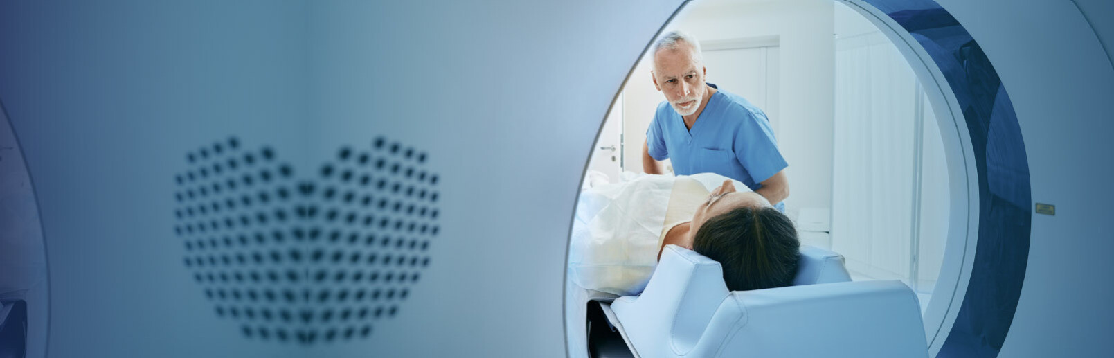
CT (computed tomography) uses highly sophisticated x-ray equipment and computer processing to obtain detailed images from different angles around the body. The introduction of CT scans in 1982 revolutionized non-invasive diagnostics by providing clear images of bone, organs, blood vessels and soft tissue. Today, technology has advanced to the point where multi-slice CT allows radiologists to view anatomy in a 3 dimensional fashion.
A CT scan is painless but it will require that you lie still for as long as five minutes. Some CT exams may require that drink a flavored oral contrast while others involve an intravenous (IV) contrast to be administered. CT studies are a useful diagnostic tool for many parts of the body including the head, neck, chest, spine, heart and numerous other areas.
With diagnostic x-ray images, our radiologists can review and interpret basic diagnostic studies for the purpose of determining if further evaluation may be necessary. Diagnostic x-ray images can assist the radiologist in front-line diagnostic evaluation and possibly provide enough information for treatment and final evaluation. Diagnostic x-ray is a very common procedure for determining common ailments from fractures to pneumonia.
Fluoroscopy is an imaging technique that uses X-rays to obtain real-time moving images of the internal structures of a patient through the use of a fluoroscope. In its simplest form, a fluoroscope consists of an X-ray source and fluorescent screen between which a patient is placed. However, modern fluoroscopes couple the screen to an X-ray image intensifier and CCD video camera allowing the images to be recorded and played on a monitor.
Mammography is the process of using low-energy X-rays to examine the human breast and is used as a diagnostic and a screening tool. The goal of mammography is the early detection of breast cancer, typically through the identification of characteristic masses and/or micro-calcifications. Because it is estimated that one out of every eight women will develop breast cancer in their lifetime, DIA offers highly accurate digital mammography and computer aided detection (CAD) along with their medical training and expertise, to increase the likelihood of early detection of breast disease.
MRI (magnetic resonance imaging) assists physicians in detecting and defining the differences between healthy, diseased or injured tissue. This technology uses magnetic fields, radio waves and complex computer processing to produce exceptionally clear images of organs, soft tissues, bone and other internal body structures for diagnostic purposes. Functional MRI was developed in 1992.
MRI exams are painless and can be completed with no exposure to radiation. Most MRI exams can be completed in an hour or less
Nuclear Medicine is a safe and painless imaging technology that uses radioactive tracers and a special camera to visualize the structure and function of an organ, tissue, bone or system of the body. Small amounts of radiation are used, and the radiation exposure from nuclear medicine procedures is comparable to that received from x-ray procedures.
PET/CT is used to help physicians more accurately diagnose and identify cancer, heart disease and brain disorders. PET/CT scans combine two scanning technologies—Positron Emission Tomography (PET) and Computed Tomography (CT)—into one procedure, producing a more accurate picture of what’s going on in the body than either scan alone. The PET scan produces images highlighting areas of high metabolic activity, and the CT scan produces pictures of the body’s internal structures. Put together, the combined image provides information about cell activity and its accurate location.
Ultrasound imaging, also called sonography, is a noninvasive exam that uses high-frequency sound waves to produce moving pictures of the inside of the body. Ultrasound exams are painless, no radiation, dyes or magnets are used and the technology is very safe. For this reason ultrasound exams are frequently used during pregnancy. Because ultrasound images are captured in real-time, they can show the structure and movement of the body's internal organs, as well as blood flowing through the vessels.